Search This Blog

Showing posts with label 17 - BLOOD RELATED. Show all posts

Wednesday, October 13, 2021

Myelofibrosis


Myelofibrosis

Symptoms & causes
Diagnosis & treatment
Doctors & departments
Care at Mayo Clinic
Print

Overview

Myelofibrosis is an uncommon type of bone marrow cancer that disrupts your body's normal production of blood cells.

Myelofibrosis causes extensive scarring in your bone marrow, leading to severe anemia that can cause weakness and fatigue. Bone marrow scarring can also cause you to have a low number of blood-clotting cells called platelets, which increases the risk of bleeding. Myelofibrosis often causes an enlarged spleen.


Myelofibrosis is considered to be a chronic leukemia — a cancer that affects the blood-forming tissues in the body. Myelofibrosis belongs to a group of diseases called myeloproliferative disorders.

Myelofibrosis can happen on its own (primary myelofibrosis) or it can develop from another bone marrow disorder (secondary myelofibrosis).

Some people with myelofibrosis have no symptoms and might not need treatment right away. Others with more-serious forms of the disease might need aggressive treatments right away. Treatment for myelofibrosis, which focuses on relieving symptoms, can involve a variety of options.

Products & Services

Book: Mayo Clinic Family Health Book, 5th EditionShow more products from Mayo Clinic

Symptoms

Myelofibrosis usually develops slowly. In its very early stages, many people don't experience signs or symptoms.

As disruption of normal blood cell production increases, signs and symptoms may include:
Feeling tired, weak or short of breath, usually because of anemia
Pain or fullness below your ribs on the left side, due to an enlarged spleen
Easy bruising
Easy bleeding
Excessive sweating during sleep (night sweats)
Fever
Bone pain
When to see a doctor

Make an appointment with your doctor if you have any persistent signs and symptoms that worry you.
Request an Appointment at Mayo Clinic
Causes

Myelofibrosis occurs when bone marrow stem cells develop changes (mutations) in their DNA. The stem cells have the ability to replicate and divide into the multiple specialized cells that make up your blood — red blood cells, white blood cells and platelets.

It's not clear what causes the genetic mutations in bone marrow stem cells.

As the mutated blood stem cells replicate and divide, they pass along the mutations to the new cells. As more and more of these mutated cells are created, they begin to have serious effects on blood production.

The end result is usually a lack of red blood cells — which causes the anemia characteristic of myelofibrosis — and an overabundance of white blood cells and varying levels of platelets. In people with myelofibrosis, the normally spongy bone marrow becomes scarred.

Several specific gene mutations have been identified in people with myelofibrosis. The most common is the Janus kinase 2 (JAK2) gene mutation. Other less common mutations include CALR and MPL. Some people with myelofibrosis don't have any identifiable gene mutations. Knowing whether these gene mutations are associated with your myelofibrosis helps determine your prognosis and your treatment.
Risk factors

Although the cause of myelofibrosis often isn't known, certain factors are known to increase your risk:
Age. Myelofibrosis can affect anyone, but it's most often diagnosed in people older than 50.
Another blood cell disorder. A small portion of people with myelofibrosis develop the condition as a complication of essential thrombocythemia or polycythemia vera.
Exposure to certain chemicals. Myelofibrosis has been linked to exposure to industrial chemicals such as toluene and benzene.
Exposure to radiation. People exposed to very high levels of radiation have an increased risk of myelofibrosis.
Complications

Complications that may result from myelofibrosis include:
Increased pressure on blood flowing into your liver. Normally, blood flow from the spleen enters your liver through a large blood vessel called the portal vein. Increased blood flow from an enlarged spleen can lead to high blood pressure in the portal vein (portal hypertension). This in turn can force excess blood into smaller veins in your stomach and esophagus, potentially causing these veins to rupture and bleed.
Pain. A severely enlarged spleen can cause abdominal pain and back pain.
Growths in other areas of your body. Formation of blood cells outside the bone marrow (extramedullary hematopoiesis) may create clumps (tumors) of developing blood cells in other areas of your body. These tumors may cause problems such as bleeding in your gastrointestinal system, coughing or spitting up of blood, compression of your spinal cord, or seizures.
Bleeding complications. As the disease progresses, your platelet count tends to drop below normal (thrombocytopenia) and platelet function becomes impaired. An insufficient number of platelets can lead to easy bleeding — an issue that you and your doctor will want to discuss if you're contemplating any type of surgical procedure.
Acute leukemia. Some people with myelofibrosis eventually develop acute myelogenous leukemia, a type of blood and bone marrow cancer that progresses rapidly.
By Mayo Clinic Staff

Myelofibrosis care at Mayo Clinic
Request an Appointment at Mayo Clinic

Monday, July 5, 2021

A Study of Patterns of Platelet Counts in Alcohol Withdrawal


A Study of Patterns of Platelet Counts in Alcohol Withdrawal

Devavrat G. Harshe, Harshal Thadasare,1 Sagar B. Karia,1 Avinash De Sousa,1 Rashmin M. Cholera,2 Sanjiv S. Kale,2 Omkar S. Mate,2 and Nilesh Shah1
Author information Copyright and License information Disclaimer


Go to:
Abstract

Aims:

This study aimed to evaluate the patterns of platelet counts during the course of alcohol withdrawal and its relationship if any with liver enzymes.

Methodology:

Forty consecutive patients, with alcohol dependence according to the Diagnostic and Statistical Manual of Mental Disorders-fourth edition, Text Revision criteria, willing for a 10-day inpatient detoxification program and presenting within 12 h of the last consumption of alcohol were recruited in the study. Details about the diagnosis and alcohol consumption patterns were assessed with a detailed psychiatric interview. After admission, routine investigations (complete blood counts [CBCs] and liver function tests) were sent and records were kept. CBC was sent for platelet counts on the 2nd, 4th, 6th, 8th, and the 10th day of alcohol withdrawal.

Results:

Nearly 40% of the patients developed delirium tremens (DT group) and rest had an uncomplicated alcohol withdrawal (ND group). Platelet counts at baseline and all the 4 days of collection were significantly lower in DT group than the ND group. Platelet counts increased gradually from baseline till 10th day of alcohol withdrawal, mean increase in platelet counts being 88.61 ± 11.60% (median: 61.11%, range [23.41–391.23%]). Platelet counts in 63% of the patients showed a drop on the 4th day of withdrawal before rising till the 10th day of alcohol withdrawal. Platelet counts were not affected by liver enzymes or other alcohol consumption patterns.

Conclusions:

Transient thrombocytopenia and reverse thrombocytosis during alcohol withdrawal are associated with an initial drop in platelet counts. The synchrony between the drop and the onset of DT needs to be evaluated.

Keywords: Delirium, reverse, thrombocytopenia, transient
Go to:

INTRODUCTION

Alcohol withdrawal syndrome (AWS) is a clinical entity which ensues after a sudden cessation of or reduction in quantity of absolute alcohol consumed daily in those who are dependent on alcohol. AWS has a self-limiting course in most cases, 5%–20% of patients however develop a complicated AWS[1] with alcohol withdrawal seizures or delirium tremens (DT).

Evidence suggests that low platelet count has a good predictive power in predicting the development of DT[2,3,4,5,6] in cases of AWS. Kim et al., 2015,[3] Berggren et al., 2009,[4] Eyer et al., 2011,[5] and Monte et al., 2009,[6] demonstrated that platelet count in patients with DT was in thrombocytopenia range (<150 × 109/L) whereas Huang et al., 2011,[2] showed a nonsignificantly lower platelet count in DT group as compared to ND group. A recent systematic review and meta-analysis[7] substantiated this observation. Most of the authors hypothesized that chronic alcohol consumption, heavy alcohol intake, recent binging, and comorbid liver cirrhosis may be the causative factors for thrombocytopenia during alcohol withdrawal.

Interestingly, with the exception of Berggren et al., 2009,[4] who showed a significantly higher aspartate transaminase (AST) levels in patients with DT compared to those without DT, neither any study nor the meta-analysis showed any significant difference between DT and non-DT groups for AST and alanine transaminase (ALT)! Thus, we planned this study to evaluate the stability of platelet count during the course of alcohol withdrawal and its relationship if any with serum liver enzymes.

Go to:
METHODOLOGY

Sample

The study was cleared by the Institutional Ethics Committee. Eighty consecutive patients diagnosed with alcohol dependence and presented within 12 h of their last drink were screened. Patients with (1) dependence on any other substance except nicotine, (2) a history of traumatic brain injury, peripheral vascular disease, past myocardial infarction and cerebrovascular accidents, comorbid psychosis and mood disorder, and alcohol withdrawal seizure during the past alcohol withdrawal, and (3) patients taking anti-platelet/anti-coagulation medications were excluded from the study. Patients willing for a 10-day inpatient detoxification program were recruited in the study after a written and informed consent (n = 44). Patients who developed alcohol withdrawal seizure during the course of alcohol withdrawal (n = 4) were excluded from the study.

Method

Patients (n = 40) were admitted after discussing the diagnosis and treatment strategy with a consultant (assistant professor and above). Routine investigations (complete blood count [CBC]; liver function tests; and renal function tests) were sent early morning on the 2nd day of alcohol withdrawal. Patients were started on tablet lorazepam (8–12 mg/day) in divided doses and injection thiamine 100 mg 12 hourly. Provision was made for (1) injectable lorazepam in cases of a seizure and DT, (2) tablet zolpidem (10 mg) for insomnia, and (3) tablet escitalopram for depressive symptoms during the course of AWS although they were not administered on any patient enrolled in the study. Patients were observed round the clock by on-duty residents, and an orientation chart was maintained. Dose of lorazepam was adjusted according to the clinical status, orientation, and higher functions and vital signs. CBC for platelet counts was sent on alternate days (day 2, day 4, day 6, day 8, and day 10) of alcohol withdrawal. Patients were shifted on oral multi-vitamin supplementation from day 5 of alcohol withdrawal. Lorazepam was tapered gradually from 6th day onward based on the clinical improvement and stopped completely by the time of discharge. Patients were prescribed appropriate medications for relapse prevention based on clinical status, affordability, and tolerability profile.

Data analysis

Data were pooled in a spreadsheet program and analyzed. Dichotomous variables were assessed with Chi-square test and 2 × 2 cross-tabs analysis. Quantitative variables (platelet counts on 5 days of blood collection) were compared with independent samples t-test. Pearson's correlation was used to study relationship between platelet counts, serum liver enzymes, and alcohol consumption details. Statistical significance was assumed at P < 0.05.

Demographic details

Sample of 40 was an all-male sample with a mean age of 38.47 ± 9.86 years (range: 21–56 years). Mean age at the onset of alcohol consumption was 21.27 ± 4.70 years. Nearly 63% of the patients (25/40) were consuming country liquor (CL) at the time of this study and the rest were consuming whiskey. Almost 40% of the patients developed DT during the course of AWS.

Platelet counts in AWS

Nearly 30% of the patients showed thrombocytopenia. Platelet counts were significantly lower in DT group than ND group, on all five occasions. Mean platelet count on day 4 was lower than that of baseline in both DT and ND groups. Mean platelet counts showed a gradual rise on days 6, 8, and 10 of alcohol withdrawal. Nearly 63% (25/40) of the patients showed a drop in platelet counts on the 4th day of withdrawal followed by a rise whereas the rest (15/40) showed a continuous rise from 2nd day in platelet counts till 10th day of alcohol withdrawal. Mean increase in the platelet counts on day 10 from baseline was 88.61 ± 11.60% (median: 61.11%; range [23.41–391.23%]). DT group showed a significantly higher increase in platelet counts as compared to ND group on the 8th (82.24 ± 56.54 [DT] vs. 38.29 ± 16.84 [ND]%, Z = −2.457, P = 0.013) and the 10th day (151.77 ± 101.77 [DT] vs. 61.54 ± 32.08 [ND]%, Z = −2.678, P = 0.007) of AWS. Incidence of the initial drop in platelet counts did not differ between DT and ND groups (68 [DT] vs. 59 [ND]%, χ2 = 0.444, P = 0.372).

Factors affecting platelet counts

Platelet counts [Table 1] did not differ significantly in groups (1) with and without elevated liver enzymes, (2) consuming CL and Indian-made foreign liquor (IMFL), and (3) with duration of alcohol dependence (DAD) <10 and >10 years. The incidence of the initial drop in platelet counts was not affected by (1) the alcoholic beverage (74 [CL] vs. 56% [IMFL], χ2 = 1.202, P = 0.241), (2) course of alcohol withdrawal (69 [DT] vs. 60 [ND]%, χ2 = 0.444, P = 0.372), (3) serum AST (64 [elevated] vs. 60 [normal], χ2 = 0.012, P = 0.597), and (4) serum ALT levels (60 [elevated] vs. 66 [normal], χ2 = 0.181, P = 0.465). Platelet counts did not show a significant correlation with liver enzymes, age, and DAD [Table 2].

Table 1


Platelet counts (×109/L) across various clinical and biochemical factors


Table 2


Platelet counts and various clinical variables

Go to:
DISCUSSION

This study observed that platelet counts are not stable during the course of alcohol withdrawal. Platelet counts show a gradual rise from baseline till the end of alcohol withdrawal, a phenomenon which has been described as reverse thrombocytosis (RT) in literature. We also found that, in a large proportion of patients, platelet counts show a drop below the baseline in the first half of alcohol withdrawal (day 4), followed by a gradual rise till the 10th day of withdrawal. Fink and Hutton, 1983 (n = 18),[8] and Mikhailidis et al., 1986 (n = 27),[9] demonstrated RT in patients with alcohol withdrawal. Both studies, however, estimated platelet counts on day 1, day 8, and then on day 15 of alcohol withdrawal and did not report a decline in platelet counts on the 4th day. The 3rd and 4th days in the course of alcohol withdrawal have significant clinical attributes. It is the time when usually AWs and DT set in during AWS. Only further research can report whether this synchrony is of any clinical significance or is mere an accidental finding.

This study also supports the existing literature in showing that low platelet count in alcohol withdrawal is not associated with elevated liver enzymes. Thus, other possible causative factors need to be explored for the transient low platelet counts in alcohol withdrawal. Not only the count, but also platelets show structural as well as functional changes during the course of alcohol withdrawal. Platelets show a (1) decrease in platelet aggregation and thromboxane A2 secretion which returns to normal within 2 weeks of abstinence from alcohol,[8,9] (2) normalization of bleeding time during 2 weeks of abstinence,[9] and (3) a rise in platelet serotonin concentration[10] during the course of AWS among many other alterations. Thus, it can be hypothesized that alcohol withdrawal is associated with an array of changes in platelet structure and function. One of these changes is a transient thrombocytopenia (TT), which reverses eventually and in some cases, reverses after an initial drop in counts below the baseline count! The TT and the RT may be attributed to an initially increased platelet agreeability as shown by Fink and Hutton, 1983.[8] Berggren et al., 2009,[4] have hinted that thrombocytopenia at the onset of alcohol withdrawal is due to the cumulative hepatotoxic effects of alcohol. We, however, could not find any correlation between liver enzymes and platelet counts at the initial and final phases of alcohol withdrawal.

Small sample size is a key limitation in this study. A significant number of patients had to be excluded from the study due to the stringent inclusion criteria such as presentation within 12 h of the last drink and willingness to stay for a 10-day inpatient detoxification program. Inpatient management was vital to ensure strict abstinence from alcohol. This study estimated platelet counts on days 2, 4, 6, 8, and 10. It is possible that there may be more changes in platelet counts on the days when platelet counts were not estimated (days 1, 3, 5, 7, and 9).
Go to:
CONCLUSIONS

Thrombocytopenia associated with alcohol withdrawal and DT is transient and is associated with RT. RT is either continuous or is associated with an initial drop, which is followed by a continuous rise in platelet counts till withdrawal subsides. Platelet counts in alcohol withdrawal are not affected by serum AST and ALT levels, type of alcoholic beverage consumed, DAD, and quantity of daily intake of absolute alcohol.

Future directions

These findings need to be validated in a larger sample size. It would also be interested to know the patterns of platelet counts in the remaining days (day 1,3,5,7,9) of alcohol withdrawal.

Financial support and sponsorship

Nil.

Conflicts of interest

There are no conflicts of interest.
Go to:
REFERENCES
1. Finn DA, Crabbe JC. Exploring alcohol withdrawal syndrome. Alcohol Health Res World. 1997;21:149–56. [PMC free article] [PubMed] [Google Scholar]
2. Huang MC, Chen CH, Liu HC, Chen CC, Ho CC, Leu SJ. Differential patterns of serum brain-derived neurotrophic factor levels in alcoholic patients with and without delirium tremens during acute withdrawal. Alcohol Clin Exp Res. 2011;35:126–31. [PubMed] [Google Scholar]
3. Kim DW, Kim HK, Bae EK, Park SH, Kim KK. Clinical predictors for delirium tremens in patients with alcohol withdrawal seizures. Am J Emerg Med. 2015;33:701–4. [PubMed] [Google Scholar]
4. Berggren U, Fahlke C, Berglund KJ, Blennow K, Zetterberg H, Balldin J. Thrombocytopenia in early alcohol withdrawal is associated with development of delirium tremens or seizures. Alcohol Alcohol. 2009;44:382–6. [PubMed] [Google Scholar]
5. Eyer F, Schuster T, Felgenhauer N, Pfab R, Strubel T, Saugel B, et al. Risk assessment of moderate to severe alcohol withdrawal – Predictors for seizures and delirium tremens in the course of withdrawal. Alcohol Alcohol. 2011;46:427–33. [PubMed] [Google Scholar]
6. Monte R, Rabuñal R, Casariego E, Bal M, Pértega S. Risk factors for delirium tremens in patients with alcohol withdrawal syndrome in a hospital setting. Eur J Intern Med. 2009;20:690–4. [PubMed] [Google Scholar]
7. Goodson CM, Clark BJ, Douglas IS. Predictors of severe alcohol withdrawal syndrome: A systematic review and meta-analysis. Alcohol Clin Exp Res. 2014;38:2664–77. [PubMed] [Google Scholar]
8. Fink R, Hutton RA. Changes in the blood platelets of alcoholics during alcohol withdrawal. J Clin Pathol. 1983;36:337–40. [PMC free article] [PubMed] [Google Scholar]
9. Mikhailidis DP, Jenkins WJ, Barradas MA, Jeremy JY, Dandona P. Platelet function defects in chronic alcoholism. Br Med J (Clin Res Ed) 1986;293:715–8. [PMC free article] [PubMed] [Google Scholar]
10. Llinás SG, Caballero AJ, Peñalver JC, Valdés R. Platelet serotonin concentration and clinical status in alcohol withdrawal syndrome, preliminary results. MEDICC Rev. 2014;16:37–42. [PubMed] [Google Scholar]

Articles from Indian Journal of Psychological Medicine are provided here courtesy of Indian Psychiatric Society South Zonal Branch


Formats:
Article |
PubReader |
ePub (beta) |
Printer Friendly |
Cite

Share
Facebook
Twitter
Google+

Save items

Add to FavoritesView more options

Similar articles in PubMed

Predictors of severe alcohol withdrawal syndrome: a systematic review and meta-analysis.[Alcohol Clin Exp Res. 2014]
Thrombocytopenia in early alcohol withdrawal is associated with development of delirium tremens or seizures.[Alcohol Alcohol. 2009]
Clinical predictors for delirium tremens in patients with alcohol withdrawal seizures.[Am J Emerg Med. 2015]
Alcohol-induced thrombocytopenia: Current review.[Alcohol. 2020]
The role of somatic disorders and physical injury in the development and course of alcohol withdrawal delirium.[Alcohol Clin Exp Res. 1999]See reviews...See all...

Links

MedGen
PubMed
Taxonomy

Recent Activity
ClearTurn Off
A Study of Patterns of Platelet Counts in Alcohol Withdrawal
Indian Journal of Psychological Medicine. Jul-Aug 2017; 39(4)441

Pharmacogenomics of Anti-platelet and Anti-coagulation Therapy
NIHPA Author Manuscripts. 2013 Jul; 15(7)
See more...


Review Exploring alcohol withdrawal syndrome.[Alcohol Health Res World. 1997]


Differential patterns of serum brain-derived neurotrophic factor levels in alcoholic patients with and without delirium tremens during acute withdrawal.[Alcohol Clin Exp Res. 2011]
Clinical predictors for delirium tremens in patients with alcohol withdrawal seizures.[Am J Emerg Med. 2015]
Thrombocytopenia in early alcohol withdrawal is associated with development of delirium tremens or seizures.[Alcohol Alcohol. 2009]
Risk assessment of moderate to severe alcohol withdrawal--predictors for seizures and delirium tremens in the course of withdrawal.[Alcohol Alcohol. 2011]
Risk factors for delirium tremens in patients with alcohol withdrawal syndrome in a hospital setting.[Eur J Intern Med. 2009]
Review Predictors of severe alcohol withdrawal syndrome: a systematic review and meta-analysis.[Alcohol Clin Exp Res. 2014]


Thrombocytopenia in early alcohol withdrawal is associated with development of delirium tremens or seizures.[Alcohol Alcohol. 2009]


Changes in the blood platelets of alcoholics during alcohol withdrawal.[J Clin Pathol. 1983]
Platelet function defects in chronic alcoholism.[Br Med J (Clin Res Ed). 1986]


Changes in the blood platelets of alcoholics during alcohol withdrawal.[J Clin Pathol. 1983]
Platelet function defects in chronic alcoholism.[Br Med J (Clin Res Ed). 1986]
Platelet serotonin concentration and clinical status in alcohol withdrawal syndrome, preliminary results.[MEDICC Rev. 2014]
Thrombocytopenia in early alcohol withdrawal is associated with development of delirium tremens or seizures.[Alcohol Alcohol. 2009]


Support CenterSupport Center

Wednesday, April 28, 2021

People with 'O' blood group less vulnerable to COVID-19:


People with 'O' blood group less vulnerable to COVID-19: Indian research body

WION Web Team
New Delhi, Delhi, India Published: Apr 28, 2021, 05:39 PM(IST)


Blood test Photograph:( Reuters )
FOLLOW US

STORY HIGHLIGHTS


The paper has also suggested that a vegetarian diet, which is rich in fibre, might also play an important role in providing immunity against the deadly virus


Council of Scientific Industrial Research (CSIR), a research institute in India, has conducted a serosurvey in India to determine which blood groups are more vulnerable to the deadly coronavirus.

The pan-India analysis has revealed that people with blood group 'O' are less susceptible to COVID-19 and people with the blood groups 'B' and 'AB' are more vulnerable to the virus.


Also read | BioNTech founder 'confident' Covid vaccine works against Indian variant

The blood group 'O' has been associated with a low positivity rate, as per the experts of CSIR, whereas 'AB' ranked highest in seropositivity, followed by the 'B' blood group.


This survey was conducted by a team of 140 doctors who assessed samples from nearly 10,427 adults from all over the country, who became subjects of this research voluntarily.

Also read | World has failed India amid record surge in COVID-19 cases, says top US disease expert Fauci

It has also been suggested in the research that smoking, which is labelled to be a deadly habit, might be acting as a line of defence as it helps increase mucous production. However, the researchers have also stressed that further study is needed into this matter and CSIR does not promote smoking in either case.

"Smoking is known to be severely detrimental to health and associated with multiple diseases and this observation should not be taken to be an endorsement, especially given that the association is not proved to be causal," the paper said.

The paper has also suggested that a vegetarian diet, which is rich in fiber, might also play an important role in providing immunity against the deadly virus.

Sunday, March 19, 2017

10 Causes of High Platelet Count - THROMBOCYTE.COM


High Platelet Count
Platelets are small, oval/spherical component of the blood that don’t have its own nucleus. These small fragments are produced in the bone marrow and plays a very important part in blood clotting.

Normal levels of platelets in the blood is about 150-350×109 in 1 mm³ blood. Platelets life span goes for only 7-10 days.

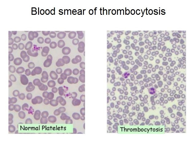
Doctors can detect thrombocytosis during routine blood tests. A display of increased level of platelets can be helpful in determining reactive thrombocytosis or thrombocythemia, which may likely cause abnormal blood clots.

What causes high platelet count? Thrombocytosis is the medical term used to describe the display of elevated platelet count. The higher end for normal platelet count range may vary from lab to lab, but is typically around 350 to 450 × 109 / L.
With the information below, we will identify further what causes platelets to be high in comparison to normal levels. There are over 10 factors actually. Also, check out the most common platelet disorders.

The causes of high platelet count or thrombocytosis can be classified as follows:
  • Physiological thrombocytosis
  • Reactive (secondary) thrombocytosis
  • Clonal (primary) thrombocytosis

I. THE PHYSIOLOGICAL THROMBOCYTOSIS CAN BE A RESULT OF:
  • Exercise (workload)
  • Stress
  • Adrenaline
II. THE REACTIVE (SECONDARY) THROMBOCYTOSIS MAY RESULT FROM:
  • Acute blood loss
  • Hemolytic anemia
  • Infection
  • Inflammatory diseases
  • Iron deficiency anemia
  • Malignant disease
  • Surgery
  • Post splenectomy / hypospleenism
  • Drug reactions (vincristine, all trans retinoic acid, cytokines, growth factors)
  • Feedback (“reactive”) thrombocytosis
  • Trauma
Infection
A series of acute and chronic infections has been associated with reactive thrombocytosis. Megakaryopoiesis is inhibited during the presence of an acute infection which may be due to a virus or bacteria. This identifies what causes a high platelet count in a patient who is experiencing either a viral or bacterial infection. Bacterial infections may be pneumonia, pyelonephritis, purulent arthritis, osteomyelitis, chronic wound infections, tuberculosis, among others. Viral infections, on the other hand, rarely reflects thrombocytosis.

Inflammatory diseases
The inflammatory diseases that could cause thrombocytosis are rheumatoid arthritis, rheumatic polymyalgia, polyarteritis nodosa, inflammatory bowel disease, nephritis and liver cirrhosis. Typically, the degree of severity of the disease condition corresponds to that of thrombocytosis. With proper treatment of the involved inflammatory disease/s, platelet count is very likely to return to normal levels.

Iron deficiency anemia
Elevated platelet count is not uncommon in patients with sideropenic anemia, specifically iron deficiency anemia. Sometimes platelet count may be greater than 1000 × 109 / L. Introducing iron replacement therapy helps the platelet count to generally return to normal within 10 days.

Malignant disease
There’s also been a described association of thrombocytosis with many neoplasms, including Hodgkin’s lymphoma, non-Hodgkin’s lymphoma, ovarian, bladder, mesothelioma, lung cancer, prostate and pancreatic cancer. About 90% of patients with reactive thrombocytosis due to a malignant disease displays a platelet count between 400 and 1000 × 109 / L.
Splenectomy

After splenectomy, a person may develop thrombocytosis that sometimes exceed 1000 × 109 / L. The removal of the spleen is what causes high platelets in some patients after the surgery. The platelet count usually returns to normal within a few weeks or months. Permanent thrombocytosis after splenectomy should give pause for thought about the existence of myeloproliferative disorder or any condition that may have developed like hemolysis or ineffective erythropoiesis.

Reactive thrombocytosis rarely causes symptoms.

BUT IF SYMPTOMS OCCUR, THEY MAY INCLUDE THE FOLLOWING:
  • Headache
  • Dizziness or lightheadedness
  • Chest pain
  • Weakness
  • Unconsciousness
  • Temporary changes in vision
  • Numbness or tingling in the hands or feet

TREATMENT OF REACTIVE THROMBOCYTOSIS
Treatment of reactive thrombocytosis is directed at the cause. If the cause is due to previous surgery or injury that have resulted to a significant loss of blood, thrombocytosis will not last long. If the cause is due to chronic infection or inflammatory disease, platelet levels may remain elevated until the condition is brought into control.

In most cases, the platelet count returns to normal after treatment of the root cause. Still yet, the removal of the spleen may cause a lifetime of thrombocytosis. In this case, the doctor may prescribe a low-dose aspirin to prevent bleeding as well as blood clotting, but which rarely occur in reactive thrombocytosis.

III. THE CLONAL OR PRIMARY THROMBOCYTOSIS MAY APPEAR BECAUSE OF:
  • AML (acute megakaryocytic leukemia)
  • Myeloproliferative disease
  • Essential thrombocytosis
  • Polycythemia vera
  • Agnogenic myeloid metaplasia
  • Chronic myeloid leukemia
  • Myelodysplastic syndrome
Chronic myeloid leukemia
Two-thirds of patients with chronic myeloid leukemia are found to have thrombocytosis.

Polycythemia vera
Increased platelet count has also been observed in approximately 66% of patients with polycythemia vera. About 5% of the patients have marked thrombocytosis (platelet count greater than 1000 x 109 / L).

Agnogenic myeloid metaplasia
Also known as idiopathic myelofibrosis, myeloid metaplasia agnogenic, is manifested by anemia and splenomegaly. A review of peripheral blood smear often detects 

Leukoerythroblastosis, described as a finding of erythrocytes in the form of tears, and immature precursors of red blood cells and leukocytes.
Thrombocytosis has been found in about 33% of patients with the condition, but in advanced stages of the disease, thrombocytopenia will usually be the significant finding.
For most myelodysplastic disorders, thrombocytosis is not typical. In fact, in most patients, it’s more often a display of normal or reduced platelet count. One form of myelodysplastic syndrome, called “5q-syndrome”, is associated with thrombocytosis in 50% of patients.

Essential thrombocytosis
Essential thrombocytosis, or primary thrombocythemia is a chronic disease characterized as a myeloproliferative neoplasm showing an enlargement in the total number of blood platelets (thrombocytes).

The exact cause of essential thrombocythemia is not fully known, but it is assumed that there is a certain degree of genetic predisposition.

Some research shows that essential thrombocythemia is diagnosed in only about 6 persons per 100,000 people in one year. Males are equally affected as females, however with the younger population, a higher percentage goes to females. This disease is more common among members of the older population, although one in five patients is younger than 40 years.

SYMPTOMS OF ESSENTIAL THROMBOCYTOSIS
Nearly every third patient at the time of diagnosis are without any signs.
Among the signs and symptoms, one of the most known insight to look for is that high platelets causes thrombosis of blood vessels. Read about the common high platelet count symptoms.

The most frequently occurring symptoms:
  • Headache is the most common neurological symptom. Others include problems with speech, dizziness, fainting, loss of vision and seizures.
High platelet count causes the fingers to likely develop pain and become affected with gangrene
  • Thrombosis of large blood vessels affect the blood vessels that supply the extremities (deep vein thrombosis)
  • It can also affect the blood vessels of the heart (coronary syndrome)
  • The gastrointestinal tract is also often found with forms of bleeding complications
  • Bleeding can also occur under the skin, gums, joints and brain tissue
  • Signs and symptoms like the occurrence of loss of appetite and body weight may also take place

DIAGNOSIS OF ESSENTIAL THROMBOCYTOSIS
Medical history, analysis of the clinical picture, and detailed overview can help in identifying reasons for high platelet count in a patient. This should come along with supplementary medical tests and examinations. The definitive diagnosis is made after laboratory procedures and measurements pertaining to platelet levels of the blood has been completed. Also, the identification of elevated platelet count causes behind the patient’s condition, comes with analysis that is carried further to associated factors like anemia and increase in leukocytes.

If the diagnostic process requires, a bone marrow sample may be necessary.

HOW TO TREAT ESSENTIAL THROMBOCYTOSIS:

The treatment of essential thrombocytosis involves administration of hydroxyurea, interferon alfa, radioactive phosphorus 32 and low-dose aspirin on a daily basis.

Furthermore, the use of Shiitake mushroom is considered as one of the best natural management options to regulate thrombocytosis. The plant’s essential oil has been known to inhibit platelet aggregation.

In emergency cases, we can use plasmapheresis.

Thursday, September 15, 2016

8 Foods That Could Help Unclog Your Arteries


Saturday, May 23, 2015

Acid/Alkaline Balance: Something Everyone Should Know


Health is truly a balance among all the systems of the body. The cells of our body are so interconnected that when you improve the balance of any one system, it improves the balance and vitality of the rest of the organ systems. For example, improving cardiovascular health improves digestive function. Improving the function of the nervous system will improve the lymphatic system, and possibly most important, when you improve the acid/alkaline balance of the body, you simultaneously improve every other system in the body at the same time.

Finding balance begins in our bloodstream, as does repairing injuries, reducing inflammation, burning fat, building strong bones (reversing osteoporosis), and the increase of energy and vitality.

In order to function properly, the blood and other body fluids must maintain a very narrow acid/alkaline balance, which is measured by the pH factor (potential hydrogen). pH ranges from 0-14 (very acidic to very alkaline).  A pH below 7.0 is acid and above 7 is alkaline. Keep in mind when and if you test your pH that there is a tenfold difference between each number. For example, a pH of 5.0 is 10 times more acidic than a pH of 6.0

Blood pH does not shift easily. The pH of our blood is between 7.25-7.45, and if the blood’s pH falls below or above that range, the body cannot function properly. A tremendous amount of energy is expended to maintain pH levels, all the while pulling from the body’s alkaline mineral reserves, causing deficiencies and health disorders.

When we maintain proper pH levels, injuries heal faster and health challenges improve more quickly because the body is oxygenated and therefore can detoxify and heal itself. If cells are energized in this way, we develop a strong immunity to diseases and a significantly lower propensity for cancer.

The best way to maintain optimal pH levels and optimal vitality is through what we eat and drink and how we react to stress. For the diet, if you follow an 80/20 rule, 80% alkaline forming foods and 20% acid, you should experience all the benefits that a body balance has to offer. Have a look at the following lists to see which foods do what and where you might be able to increase your alkaline-forming foods. Keep in mind that the foods must be organic because ALL pesticides are very acidic.

ALKALINE FORMING FOODS:

DAIRY: acidophilus, whey, kefir/yogurt

FRUITS: apples, apricots, avocados, bananas, berries, cantaloupes, cherries, currants, dates, figs, grapes, grapefruits, guavas, lemons, limes, mangoes, melons, nectarines, oranges, papayas, passion fruits, peaches, pears, persimmons, pineapples, raisins, strawberries, tangerines

VEGGIES: bamboo shoots, green beans, lima beans, string beans, sprouts, beets, broccoli, cabbage, carrots, celery, cauliflower, chard, chicory, chives, collard greens, cucumber, dandelion greens, dill, dulse, eggplant, endive, escarole, kale, garlic, leeks, legumes, lettuce, okra, onions, parsley, parsnips, sweet potato/yam, bell peppers, white potatoes, pumpkin, radish, rutabaga, turnips, watercress

MEAT: No meat is alkaline

NUTS: almonds, chestnuts, coconuts

MISC: ginger, honey, kelp, alfalfa, clover, mint, sage, green tea, quinoa, flaxseed, pumpkin seeds, all seaweed/sea veggies

MINERALS: calcium, magnesium, potassium, manganese


ACIDIFYING FOODS & FACTORS

GRAINS: all white flour products, buckwheat, wheat, corn, barley, oats, rye

DAIRY: butter, eggs, cheese, cottage cheese, cream, ice cream, custards, milk

FRUITS: jams/anything preserved, cranberries, pomegranates, olives

VEGGIES: artichokes, asparagus, garbanzo beans,

MEAT: All

NUTS: peanuts, pistachios, walnuts, macadamia

MISC: alcohol, brine, coffee, cocoa/chocolate, candy, many dressings (because of the vinegar), drugs, jams/jellies, mayo, some spices, soda,

ALSO lack of sleep, stress, worry


Saturday, May 2, 2015

The 11 Health Benefits of Whisky


The 11 Health Benefits of Whisky

Whisky is one of the most popular alcoholic beverages in the world, thanks to its distinct flavors and the fact that it’s the least likely to give you a hangover. Add to that the fact that whisky also holds several medicinal property, and you got yourself an obvious success story.

Please remember that whisky is alcoholic, so drink it responsibly and as always - in moderation.


A Brief History

The first confirmed accounts of whisky distillation are from the early 15th century, when the Irish and Scottish distilled it, calling it “Aqua Vitae” (Water of Life / Lively Water), for medicinal properties. 


During the American Revolution, it was used as currency and George Washington even owned a distillery in Mount Vernon. In the Prohibition era (1920-1933), whisky was the only alcohol permitted for consumption with a doctor’s prescription.


The Health Benefits of Whiskey:

1. Memory Boost: Whisky contains antioxidants that help improve the health of the brain, additionally, alcohol boosts blood circulation, both of which contribute to your memory. In addition, the Ethanol in whisky helps your neurons function properly, which also aids with memory.

2. Stress Relief: In moderation, whisky can reduce stress and calm the nerves. The combination of slowing down  brain activity and increased circulation, which helps provide the body with oxygenated blood, are essential for achieving calmness.


3. Fight Weight Gain: Compared to its counterparts, whisky is a low-calorie alcohol, free of fat and cholesterol. If you’re on a diet but still want a drink – it’s your best choice.

4. Reduce the Risk of a Stroke: Whisky prevents cholesterol from accumulating in the cardiovascular system and can help remove excess cholesterol from the body. It also relaxes the walls of the arteries, reducing the risk of obstruction. All of these factors help reduce the risk of stroke considerably.

5. Reduce the Risk of Cancer: Whisky contains an antioxidant called ‘ellagic acid’, an acid that stops DNA from coming in contact with cancer-causing compounds, such as nitrosamines and polycyclic aromatic hydrocarbons.. It is also said to help protect the body from damage during chemotherapy.

6. Helps with Digestion: For centuries, whisky was considered a digestive aid, drunk after a heavy meal. Whisky's composition and high alcohol percentage also makes it an effective appetite suppressant.

7. Live Longer: The antioxidants in whisky help fight free radicals – the number one cause of aging, as well as prevent various diseases. This double-whammy helps your body live a longer, healthier life.

8. Diabetic-Friendly: Containing zero carbs, it won’t affect blood sugar levels, making it the number one choice for diabetics.

9. Improve Your Heart's Health: Drinking whisky actually helps your heart stay healthy, similarly to red wine. It reduces the risk of blood clots, thus it can prevent strokes and heart attacks. The antioxidants in the whisky also inhibits the oxidation of low density lipoprotein - a main factor in heart disease.

10. Improve the Health of Your Brain: A 2003 study found that, thanks to the antioxidant qualities of the ellagic acid, moderate consumption of whisky reduces the risk of Alzheimer’s and dementia, as well as improve cognitive functions. Basically, one drink a day will keep the doctor away.

11. Prevent & Treat Cold and Flu: Whisky is known for its positive effects towards allergies and colds. It’s an effective cough syrup for people suffering from an itchy throat, and the alcohol helps kill bacteria in the throat. The best results are achieved by adding a little bit of whisky to a cup of hot water and lemon.

Sunday, August 17, 2014

10 Foods to Help Prevent Clogged Arteries

AUGUST 11, 2013 by DR. NIMA SHEI

10 Foods to Help Prevent Clogged Arteries

10 Foods to Help Prevent Clogged Arteries


Garlic
Since ancient times garlic has been used to treat heart disease and hypertension. According to a study published in preventive medicine, garlic inhibits coronary artery calcification which serves as a marker for plaque formation.

Grapes
Grapes are rich in flavonoids, quercetin, and resveratrol. These flavonoids have been found to prevent the oxidation of bad cholesterol that leads to the formation of plaque in artery walls. They also lower the risk of developing blood clots that can lead to heart attacks.

Spinach
Spinach is rich in potassium and folic acids, both of which act as a defense against high blood pressure. Spinach is also rich in lutein, a plant carotenoid which not only protects against age related macular degeneration but also prevents heart attacks by keeping arteries free from cholesterol build up.

Fish
A study conducted by researchers at Southampton University found that Omega 3 oils, present in fishes like tuna and salmon, stop the build up of fatty deposits in the arteries. These fatty acids prevent clots from forming and cholesterol from becoming oxidized.

Olive oil
Only oxidized cholesterol is able to stick to your artery walls and form plaques. Monounsaturated fats present in olive oil when mixed with molecules of bad cholesterol become less likely to be oxidized.

Tomatoes
According to a study conducted by Korean researchers, lycopene, a compound present in tomatoes that gives them their color, helps in prevent hardening of the arteries. Researchers found that women with the highest lycopene levels in their blood had the least stiffness in their arteries.

Pomegranates
Pomegranate juice not only appears to prevent hardening of the arteries by reducing blood vessel damage, but may also reverse the progression of this disease. Pomegranate fruit and its juice are high in antioxidant content, which may help fight hardening of the arteries.

Kiwi and Cantaloupe
These antioxidant-rich foods work by reducing toxic LDL cholesterol, which is formed by a rusting process in your arteries. They can help stop the rust in its tracks and even prevent it from spreading. Eat one cup of cantaloupe or one kiwi per day to unclog your arteries.

Cranberry juice
This juice strains the fat out of arteries. Instead of having fat build up in your blood vessels, this juice boosts your cell’s ability to absorb the fat and use it for production of energy. Drink three glasses of this juice every week to unclog your arteries.

Oats
Thanks to the soluble fiber in oats, cholesterol doesn’t stand a chance. This fiber attaches itself to cholesterol and carries it right out of your system. Some studies have shown that eating just 1 1/2 to 2 1/2 cups of cooked oat-bran cereal daily can lower cholesterol levels by almost 20%.

10 Foods to Help Prevent Clogged Arteries
By Positivemed Team
Edited By: Stephanie Dawson