Search This Blog

Showing posts with label 15 - STOMACH RELATED. Show all posts

Thursday, December 23, 2021

Gastrointestinal bleeding - Mayo Clinic


Gastrointestinal bleeding

Symptoms & causes
Diagnosis & treatment
Doctors & departments
Care at Mayo Clinic
Print

Diagnosis
EndoscopyOpen pop-up dialog box

Your doctor will take a medical history, including a history of previous bleeding, conduct a physical exam and possibly order tests. Tests might include:
Blood tests. You may need a complete blood count, a test to see how fast your blood clots, a platelet count and liver function tests.

Stool tests. Analyzing your stool can help determine the cause of occult bleeding.

Nasogastric lavage. A tube is passed through your nose into your stomach to remove your stomach contents. This might help determine the source of your bleed.

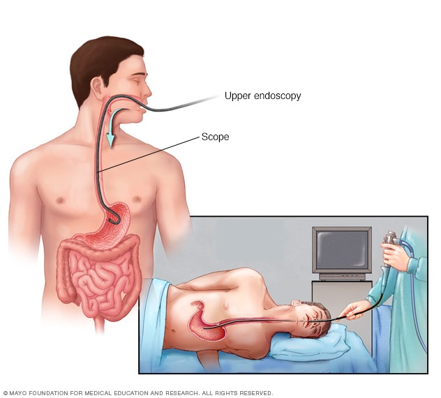
Upper endoscopy. This procedure uses a tiny camera on the end of a long tube, which is passed through your mouth to enable your doctor to examine your upper gastrointestinal tract.

Colonoscopy. This procedure uses a tiny camera on the end of a long tube, which is passed through your rectum to enable your doctor to examine your large intestine and rectum.

Capsule endoscopy.
In this procedure, you swallow a vitamin-size capsule with a tiny camera inside. The capsule travels through your digestive tract taking thousands of pictures that are sent to a recorder you wear on a belt around your waist. This enables your doctor to see inside your small intestine.

Flexible sigmoidoscopy. A tube with a light and camera is placed in your rectum to look at your rectum and the last part of the large intestine that leads to your rectum (sigmoid colon).

Balloon-assisted enteroscopy. A specialized scope inspects parts of your small intestine that other tests using an endoscope can't reach. Sometimes, the source of bleeding can be controlled or treated during this test.

 A contrast dye is injected into an artery, and a series of X-rays are taken to look for and treat bleeding vessels or other abnormalities.

Imaging tests. A variety of other imaging tests, such as an abdominal CT scan, might be used to find the source of the bleed.


If your GI bleeding is severe, and noninvasive tests can't find the source, you might need surgery so that doctors can view the entire small intestine. Fortunately, this is rare.

Care at Mayo Clinic

Sunday, October 10, 2021

What Your Farts Are Trying To Tell You


What Your Farts Are Trying To Tell You


Today’s blog is republished from my friends at TheAlternativeDaily, a leading publisher of daily alternative health tips that I personally read every day…

By Krista Hillis, TheAlternativeDaily.com

There are some things that remain unchanged from child to adulthood — and giggling when you toot is one of them. Whether you blame it on the dog or your husband, passing gas is a normal bodily function. However, is it possible to fart too often? Are your daily farts trying to tell you something? If you have noticed that you’re overly gassy lately, here’s what you need to know.

What are farts, anyway?

The average person farts nearly every day, so what is it that we’re passing? Also known as “flatulence,” a fart is simply an internal build-up of gas. More specifically, farts are made up of nitrogen, carbon dioxide, oxygen, methane and hydrogen. What a lovely little gaseous cocktail, eh

Although these gases are formed during the process of respiration and digestion, the true cause of excessive gas will depend on personal circumstances. For the most part, farts are silent and odorless. It’s estimated that only around one percent of farts produce a foul smell.

No need to be embarrassed. It’s quite natural and, in many cases, healthy! In fact, a healthy person may pass gas around 15 to 20 times per day. But is that a “normal” standard?
What causes farting?


There are a number of reasons why gas gets trapped in the body, including swallowing air, constipation and changes in microflora. When someone begins to experience an excessive buildup of gas, this may mean:
You’re suffering from intolerances or food allergies

You’re experiencing bacterial overgrowth
You are constipated

You are experiencing symptoms of IBS, Crohn’s, etc.

Fermentation is occurring within the gut

More often than not, farting is harmless. It is a normal metabolic function. And for those who consume plenty of fiber-rich foods, increased flatulence may indicate a diet that promotes positive health. Take beans, for example, a food that’s known to cause gas. This is due to the type of carbohydrates which ferment in the gut.

In that sense, I’m not going to throw out a “magic” number. How many times you fart during the day may vary. Do not focus too much on how often you’re farting, but why you are passing more gas than usual. What other symptoms, if any, have you noticed?

Am I an excessive farter?

If you have noticed that you’re overly gassy lately, start to keep track. How many times did you break wind throughout the day? What did you eat? Start to document these key pieces of information.

Although tooting more than 23 to 25 times daily is considered to be more than normal, if you’re farting more than that, there’s still no need to panic. A good rule of thumb is this: if excessive gas and bloating affect your quality of life, then it’s best to get a professional opinion.

In many cases, it’s something as simple as lactose intolerance. If you’re suffering from excessive gas and bloating, however, you may want to focus on the following possibilities:

1. You’re a fast eater


Do your loved ones often tell you to slow down when you’re eating? Do you eat quickly when you’re on-the-go? Perhaps you chew a lot of gum? Either way, when you eat rapidly, you tend to swallow more air. Once that air (made up of various gases) gets into your body, it has to come out somehow. If you don’t burp it up, you may find that it’s coming out the other end.

2. Your gut is imbalanced

As mentioned, a small percentage of farts are those stinky bombs that make this act so embarrassing. You would think that rotten farts indicate something is the matter, but this isn’t necessarily the case. Generally speaking, stinky gas is generally caused by the breakdown of sulfur.

Since there are many healthy foods that contain sulfur, including broccoli and beans, stinky gas does not automatically indicate poor health. With that being said, if you notice that the stench of your farts is overly awful — and your diet is less than ideal — this could mean you’re suffering from an underlying condition.

If you’re farting often, you may think that you’re unhealthy. But the truth is, passing gas can mean that your gut is being properly fed. The healthy microbes that live in our gut, are known to promote a wide range of health benefits. In order for these microbes to get nutrients, we must eat foods that cause gas.

When there’s undigested food in the large intestine, including fiber and other hard-to-digest carbs, these microbes get to work. In turn, more gas is created — as well as short-chain fatty acids, which promote the growth of other beneficial bacteria.

Farting is healthy, just not too much

I think this is important to mention, as it’s often recommended that we eat more probiotics. However, fiber is equally critical. Based on a recent study, published in Cell, researchers found that microbes need fiber in order to maintain optimal health. When they do not get natural fiber as a food source, they can actually begin to eat the layer of mucus that lines the gut — potentially leading to infection.

So, passing wind while consuming a healthy diet is fairly normal — but excessive gas could also mean you’re suffering from SIBO, or small intestinal bacterial overgrowth. Normally, the majority of bacteria is found inside the colon, but in this case, bacteria begins to invade the small intestine.

In turn, individuals experience poor nutrient absorption and symptoms often associated with IBS (irritable bowel syndrome). Along with increased flatulence, you may also experience bloating, fatigue, skin rashes and weight loss. If this is the case, you will need to address your current diet. Implement herbal remedies, including the use of lemon balm and oregano oil.

3. You’re sensitive to gluten or dairy


Even if you have been eating dairy your whole life without any noticeable issues, your system can change as you age. Over the course of time, your body may no longer produce enough lactase. The same is true for those who can not properly break down gluten — which is the case among those with celiac disease.

Whenever you believe something in your diet is to blame for any abnormal symptoms, including excessive flatulence, it’s best to try an elimination diet. Stop eating all dairy, for instance, and then see how you feel. Continue this process, focusing on FODMAP carbohydrates, including wheat, dairy, onions, garlic, legumes and stone fruit (cherries and peaches).

Although many FODMAP carbohydrates are healthy, they can be hard to break down. Each individual is different, so it’s best to keep a food journal and identify key triggers. Once you better understand the cause, you can create a more effective action plan.

Listen to your body

From hormonal changes to constipation, there are a number of reasons why you may be tootin’ more often. As mentioned, unless you fart a lot (30 or more times daily), or you’re finding that excessive flatulence is affecting your quality of life, your body is more than likely just doin’ its thing.

Like all symptoms, your body is trying to communicate with you. The more in-tune you are, the easier it will be to maintain positive health. So, listen to your body. In this case, your farts will do all the talking.

The original source of this article is found here

“Speaking of digestive issues…”

This “pooping trick” helps people slim down fast after 40

One of the reasons why losing weight after 40 is a struggle has NOTHING to do with your diet, your metabolism, or even exercising…

And everything to do with keeping your bowels active.

If you’re having pooping problems, your body isn’t digesting food and eliminating toxins properly.

So your bowels get clogged, leaving you with up to 10 pounds of toxic poop stuck in your gut.

The good news is, Stanford researchers have found a way to jumpstart the bowels and release up to 10 pounds of stuck poop to eliminate bloat and shrink your belly.

It’s already helping thousands of people lose weight and stay slim with LESS effort…

> Discover the “pooping trick” people over 40 are using to slim down fast.

Sunday, August 1, 2021

Esophageal Varices


Esophageal varices

Symptoms & causes
Diagnosis & treatment
Doctors & departments
Print

Diagnosis

Endoscopy
Open pop-up dialog box

If you have cirrhosis, your doctor should screen you for esophageal varices when you're diagnosed. How often you'll undergo screening tests depends on your condition. Main tests used to diagnose esophageal varices are:


Endoscopic exam. A procedure called upper gastrointestinal endoscopy is the preferred method of screening for esophageal varices. Your doctor inserts a thin, flexible, lighted tube (endoscope) through your mouth and into your esophagus, stomach and the beginning of your small intestine (duodenum).


The doctor will look for dilated veins, measure them, if found, and check for red streaks and red spots, which usually indicate a significant risk of bleeding. Treatment can be performed during the exam.

Imaging tests. Both abdominal CT scans and Doppler ultrasounds of the splenic and portal veins can suggest the presence of esophageal varices. An ultrasound test called transient elastography that measures scarring in the liver can help your doctor determine if you have portal hypertension, which may lead to esophageal varices.

Capsule endoscopy
. In this test, you swallow a vitamin-sized capsule containing a tiny camera, which takes pictures of the esophagus as it goes through your digestive tract. This might be an option for people who are unable or unwilling to have an endoscopic exam. 

This technology is more expensive than regular endoscopy and not as available. Capsule endoscopy can only help find esophageal varices and does not treat them.

More Information

Capsule endoscopy
CT scan
MRI

Treatment

The primary aim in treating esophageal varices is to prevent bleeding. Bleeding esophageal varices are life-threatening. If bleeding occurs, treatments are available to try to stop the bleeding.

Treatment to prevent bleeding


Treatments to lower blood pressure in the portal vein may reduce the risk of bleeding esophageal varices. 

Treatments may include:

Medications to reduce pressure in the portal vein. A type of blood pressure drug called a beta blocker may help reduce blood pressure in your portal vein, decreasing the likelihood of bleeding. These medications include propranolol (Inderal, Innopran XL) and nadolol (Corgard).


Using elastic bands to tie off bleeding veins. If your esophageal varices appear to have a high risk of bleeding, or if you've had bleeding from varices before, your doctor might recommend a procedure called endoscopic band ligation.

Using an endoscope, the doctor uses suction to pull the varices into a chamber at the end of the scope and wraps them with an elastic band, which essentially "strangles" the veins so they can't bleed. Endoscopic band ligation carries a small risk of complications, such as bleeding and scarring of the esophagus.
Treatment if you're bleeding

Bleeding esophageal varices are life-threatening, and immediate treatment is essential. Treatments used to stop bleeding and reverse the effects of blood loss include:

Using elastic bands to tie off bleeding veins. Your doctor may wrap elastic bands around the esophageal varices during an endoscopy.

Medications to slow blood flow into the portal vein. Drugs such as octreotide (Sandostatin) and vasopressin (Vasostrict) slow the flow of blood to the portal vein. The drug is usually continued for up to five days after a bleeding episode.


Diverting blood flow away from the portal vein. If medication and endoscopy treatments don't stop the bleeding, your doctor might recommend a procedure called transjugular intrahepatic portosystemic shunt (TIPS).

The shunt is an opening that is created between the portal vein and the hepatic vein, which carries blood from your liver to your heart. The shunt reduces pressure in the portal vein and often stops bleeding from esophageal varices.

But TIPS can cause serious complications, including liver failure and mental confusion, which can develop when toxins that the liver normally would filter are passed through the shunt directly into the bloodstream.

TIPS is mainly used when all other treatments have failed or as a temporary measure in people awaiting a liver transplant.


Placing pressure on varices to stop bleeding. If medication and endoscopy treatments don't work, doctors may try to stop bleeding by applying pressure to the esophageal varices. One way to temporarily stop bleeding is by inflating a balloon to put pressure on the varices for up to 24 hours, a procedure called balloon tamponade. Balloon tamponade is a temporary measure before other treatments can be performed, such as TIPS.

This procedure carries a high risk of bleeding recurrence after the balloon is deflated. Balloon tamponade may also cause serious complications, including a rupture in the esophagus, which can lead to death.
Restoring blood volume. You might be given a transfusion to replace lost blood and a clotting factor to stop bleeding.
Preventing infection. There is an increased risk of infection with bleeding, so you'll likely be given an antibiotic to prevent infection.
Replacing the diseased liver with a healthy one. Liver transplant is an option for people with severe liver disease or those who experience recurrent bleeding of esophageal varices. Although liver transplantation is often successful, the number of people awaiting transplants far outnumbers the available organs.
Re-bleeding

There is a high risk that bleeding will recur in people who've had bleeding from esophageal varices. Beta blockers and endoscopic band ligation are the recommended treatments to help prevent re-bleeding.

After initial banding treatment, your doctor will repeat your upper endoscopy at regular intervals and apply more bands if necessary until the esophageal varices are gone or small enough to reduce the risk of further bleeding.
Potential future treatment

Doctors are exploring an experimental emergency therapy to stop bleeding from esophageal varices that involves spraying an adhesive powder. The hemostatic powder is administered through a catheter during an endoscopy. When sprayed on the esophagus, hemostatic powder sticks to the varices and may stop bleeding.

Another potential way to stop bleeding when all other measures fail is to use self-expanding metal stents (SEMS). SEMS can be placed during an endoscopy and stop bleeding by placing pressure on the bleeding esophageal varices.

However, SEMS could damage tissue and can migrate after being placed. The stent should be removed within seven days and bleeding could recur. This option is experimental and isn't yet widely available.

Esophageal varices

Esophageal varices

Esophageal varices
Open pop-up dialog box

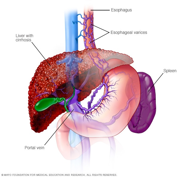
Esophageal varices are abnormal, enlarged veins in the tube that connects the throat and stomach (esophagus). This condition occurs most often in people with serious liver diseases.

Esophageal varices develop when normal blood flow to the liver is blocked by a clot or scar tissue in the liver. To go around the blockages, blood flows into smaller blood vessels that aren't designed to carry large volumes of blood. The vessels can leak blood or even rupture, causing life-threatening bleeding.


A number of drugs and medical procedures can help prevent or stop bleeding from esophageal varices.

Products & Services

Book: Mayo Clinic on Digestive Health

Symptoms

Esophageal varices usually don't cause signs and symptoms unless they bleed. Signs and symptoms of bleeding esophageal varices include:
Vomiting large amounts of blood
Black, tarry or bloody stools
Lightheadedness
Loss of consciousness in severe cases

Your doctor might suspect esophageal varices if you have signs of liver disease, including:
Yellow coloration of your skin and eyes (jaundice)
Easy bleeding or bruising
Fluid buildup in your abdomen (ascites)
When to see a doctor

Make an appointment with your doctor if you have signs or symptoms that worry you. If you've been diagnosed with liver disease, ask your doctor about your risk of esophageal varices and what you can do to reduce your risk. Also ask your doctor whether you should get a procedure to check for esophageal varices.

If you've been diagnosed with esophageal varices, your doctor is likely to instruct you to watch for signs of bleeding. Bleeding esophageal varices are an emergency. Call 911 or your local emergency services right away if you have bloody vomit or bloody stools.
Request an Appointment at Mayo Clinic
Causes

Esophageal varices sometimes form when blood flow to your liver is blocked, most often by scar tissue in the liver caused by liver disease. The blood flow begins to back up, increasing pressure within the large vein (portal vein) that carries blood to your liver.

This increased pressure (portal hypertension) forces the blood to seek other pathways through smaller veins, such as those in the lowest part of the esophagus. These thin-walled veins balloon with the added blood. Sometimes the veins rupture and bleed.

Causes of esophageal varices include:
Severe liver scarring (cirrhosis). A number of liver diseases — including hepatitis infection, alcoholic liver disease, fatty liver disease and a bile duct disorder called primary biliary cirrhosis — can result in cirrhosis.
Blood clot (thrombosis). A blood clot in the portal vein or in a vein that feeds into the portal vein (splenic vein) can cause esophageal varices.
Parasitic infection. Schistosomiasis is a parasitic infection found in parts of Africa, South America, the Caribbean, the Middle East and East Asia. The parasite can damage the liver, as well as the lungs, intestine, bladder and other organs.
Risk factors

Although many people with advanced liver disease develop esophageal varices, most won't have bleeding. Esophageal varices are more likely to bleed if you have:
High portal vein pressure. The risk of bleeding increases as the pressure in the portal vein increases (portal hypertension).
Large varices. The larger the esophageal varices, the more likely they are to bleed.
Red marks on the varices. When viewed through a thin, flexible tube (endoscope) passed down your throat, some esophageal varices show long, red streaks or red spots. These marks indicate a high risk of bleeding.
Severe cirrhosis or liver failure. Most often, the more severe your liver disease, the more likely esophageal varices are to bleed.
Continued alcohol use. Your risk of variceal bleeding is far greater if you continue to drink than if you stop, especially if your disease is alcohol related.

If you've had bleeding from esophageal varices before, you're more likely to have varices that bleed again.
Complications

The most serious complication of esophageal varices is bleeding. Once you've had a bleeding episode, your risk of another bleeding episode greatly increases. If you lose enough blood, you can go into shock, which can lead to death.
Prevention

Currently, no treatment can prevent the development of esophageal varices in people with cirrhosis. While beta blocker drugs are effective in preventing bleeding in many people who have esophageal varices, they don't prevent esophageal varices from forming.

If you've been diagnosed with liver disease, ask your doctor about strategies to avoid liver disease complications. To keep your liver healthy:
Don't drink alcohol. People with liver disease are often advised to stop drinking alcohol, since the liver processes alcohol. Drinking alcohol may stress an already vulnerable liver.
Eat a healthy diet. Choose a plant-based diet that's full of fruits and vegetables. Select whole grains and lean sources of protein. Reduce the amount of fatty and fried foods you eat.
Maintain a healthy weight. An excess amount of body fat can damage your liver. Obesity is associated with a greater risk of complications of cirrhosis. Lose weight if you are obese or overweight.
Use chemicals sparingly and carefully. Follow the directions on household chemicals, such as cleaning supplies and insect sprays. If you work around chemicals, follow all safety precautions. Your liver removes toxins from your body, so give it a break by limiting the amount of toxins it must process.
Reduce your risk of hepatitis. Sharing needles and having unprotected sex can increase your risk of hepatitis B and C. Protect yourself by abstaining from sex or using a condom if you choose to have sex. Get tested for exposure to hepatitis A, B and C, since infection can make your liver disease worse. Also ask your doctor whether you should be vaccinated for hepatitis A and hepatitis B.


By Mayo Clinic Staff

Sunday, February 7, 2021

Oesophagal Strictures,

What to Do If You Get Food Stuck in Your Throat

Seek emergency medical care
Home remedies
Medical treatment
Takeaway and prevention


We include products we think are useful for our readers. If you buy through links on this page, we may earn a small commission. Here’s our process.

Overview

Swallowing is a complex process. When you eat, around 50 pairs of muscles and many nerves work together to move food from your mouth to your stomach. It’s not uncommon for something to go wrong during this process, making it feel like you have food stuck in your throat.

When you take a bite of solid food, a three-step process begins:

You prepare the food to be swallowed by chewing it. This process allows the food to mix with saliva, and transforms it into a moistened puree.
Your swallowing reflex is triggered as your tongue pushes the food to the back of your throat. During this phase, your windpipe closes tightly and your breathing stops. This prevents food from going down the wrong pipe.

The food enters your esophagus and travels down into your stomach.

When it feels like something didn’t go all the way down, it’s usually because it’s stuck in your esophagus. Your breathing isn’t affected when this happens because the food has already cleared your windpipe. However, you may cough or gag.

Symptoms of food stuck in your esophagus develop immediately after it happens. It’s not uncommon to have severe chest pain. You may also experience excessive drooling. But there are often ways to resolve the issue at home.

When to seek emergency medical care

Thousands of people die from choking every year. It’s particularly common among young children and adults over the age of 74. Choking happens when food or a foreign object gets stuck in your throat or windpipe, blocking the flow of air.

When someone is choking, they:
are unable to talk
have difficulty breathing or noisy breathing
make squeaky sounds when trying to breathe
cough, forcefully or weakly
become flushed, then turn pale or bluish
lose consciousness

Choking is a life-threatening emergency. If you or a loved one experiences these symptoms, call your local emergency services and perform rescue techniques such as the Heimlich maneuver or chest compressions immediately.

Ways to remove food stuck in throat

The following techniques may assist you in removing food that’s become lodged in your esophagus.

The ‘Coca-Cola’ trick

Research suggestsTrusted Source that drinking a can of Coke, or another carbonated beverage, can help dislodge food stuck in the esophagus. Doctors and emergency workers often utilize this simple technique to break up food.

Although they don’t know exactly how it works, doctors believeTrusted Source that the carbon dioxide gas in soda helps disintegrate the food. It’s also thought that some of the soda gets into the stomach, which then releases gas. The pressure of the gas can dislodge the stuck food.

Try a few cans of diet soda or seltzer water at home immediately after noticing the stuck food.

Purchase seltzer water online.

Simethicone

Over-the-counter medications designed to treat gas pain may help dislodge food stuck in the esophagus. In the same way as carbonated sodas, medications containing simethicone (Gas-X) make it easier for your stomach to produce gas. This gas increases the pressure in your esophagus and can push the food loose.

Follow the standard dosing recommendation on the package.

Shop for simethicone medications.

Water

A few big sips of water may help you wash down the food stuck in your esophagus. Normally, your saliva provides enough lubrication to help food slide easily down the esophagus. If your food wasn’t chewed properly, it may be too dry. Repeated sips of water may moisten the stuck food, making it go down more easily.

A moist piece of food

It may feel uncomfortable to swallow something else, but sometimes one food can help push another down. Try dipping a piece of bread in some water or milk to soften it, and take a few small bites.

Another effective option may be to take a bite of banana, a naturally soft food.

Alka-Seltzer or baking soda

An effervescent drug like Alka-Seltzer may help break down food that’s stuck in the throat. Effervescent drugs dissolve when mixed with a liquid. Similar to soda, the bubbles they produce when dissolving may help disintegrate the food and produce pressure that can dislodge it.

Find Alka-Seltzer online.

If you don’t have Alka-Seltzer, you can try mixing some baking soda, or sodium bicarbonate, with water. This may help dislodge food in the same way.

Shop for sodium bicarbonate.

Butter

Sometimes the esophagus needs an extra bit of lubrication. As unpleasant as it may sound, it may help to eat a tablespoon of butter. This can sometimes help moisten the lining of the esophagus and make it easier for the stuck food to move down into your stomach.
Wait it out

Food that gets stuck in the throat usually passes on its own, given some time. Give your body a chance to do its thing.

HEALTHLINE NEWSLETTER
Get our weekly COPD email

To help you meet the challenges of living with COPD, we’ll send you support, inspiration, and advice for both patients and caregivers.
Enter your emailSIGN UP NOW
Your privacy is important to us


Getting help from your doctor

If you’re unable to swallow your saliva and are experiencing distress, go to your local emergency room as soon as possible. If you’re not in distress but the food is still stuck, you can have an endoscopic procedure to remove the food within 24 hoursTrusted Source. After that, there’s risk of damage to the lining of your esophagus. Some doctors recommend coming in after 6 to 12 hoursTrusted Source to reduce the likelihood of damage and make the extraction easier.

During an endoscopic procedure, your doctor can identify any possible underlying causes. If you frequently get food stuck in your throat, you should consult a doctor. One of the most common problems is a narrowing of the esophagus caused by the buildup of scar tissue, or esophageal stricture. A specialist can treat esophageal stricture by placing a stent or performing a dilation procedure.

The takeaway

Getting food stuck in your throat can be frustrating and painful. If this occurs frequently, talk to your doctor about possible underlying causes. Otherwise, you may be able to avoid a trip to the emergency room by treating yourself at home with carbonated beverages or other remedies.

In the future, be particularly careful when eating meat, as it’s the most common culprit. Avoid eating too quickly, take small bites, and avoid eating while intoxicated.


FEEDBACK:

Medically reviewed by Suzanne Falck, M.D., FACP — Written by Corinne O'Keefe Osborn — Updated on March 7, 2019


6 Natural Remedies for Wheezing


Herbs and Supplements for COPD (Chronic Bronchitis and Emphysema)


Asthma and Your Diet: What to Eat and What to Avoid


Benign Esophageal Stricture

Causes
Symptoms
Complications
Diagnosis
Treatment
Outlook
Prevention


We include products we think are useful for our readers. If you buy through links on this page, we may earn a small commission. Here’s our process.


What is benign esophageal stricture?

Benign esophageal stricture describes a narrowing or tightening of the esophagus. The esophagus is the tube that brings food and liquids from your mouth to your stomach. “Benign” means it’s not cancerous.

Benign esophageal stricture typically occurs when stomach acid and other irritants damage the lining of the esophagus over time. This leads to inflammation (esophagitis) and scar tissue, which causes the esophagus to narrow.

Although benign esophageal stricture isn’t a sign of cancer, the condition can cause several problems. Narrowing of the esophagus may make it difficult to swallow. This increases the risk of choking. It can also lead to complete obstruction of the esophagus. This can prevent food and fluids from reaching the stomach.


What causes benign esophageal stricture?

Benign esophageal stricture can happen when scar tissue forms in the esophagus. This is often the result of damage to the esophagus. The most common cause of damage is gastroesophageal reflux disease (GERD), also known as acid reflux.

GERD occurs when the lower esophageal sphincter (LES) doesn’t close or tighten properly. The LES is the muscle between the esophagus and the stomach. It normally opens for a short amount of time when you swallow. Stomach acid can flow back up into the esophagus when it doesn’t close completely. This creates a burning sensation in the lower chest known as heartburn.

Frequent exposure to harmful stomach acid can cause scar tissue to form. Eventually, the esophagus will narrow.

Other causes of benign esophageal stricture include:

radiation therapy to the chest or neck
accidental swallowing of an acidic or corrosive substance (such as batteries or household cleaners)
extended use of a nasogastric tube (a special tube that carries food and medicine to the stomach through the nose)
esophageal damage caused by an endoscope (a thin, flexible tube used to look inside a body cavity or organ)
treatment of esophageal varices (enlarged veins in the esophagus that can rupture and cause severe bleeding)

Symptoms of benign esophageal stricture

Typical symptoms of benign esophageal stricture include:
difficult or painful swallowing
unintended weight loss
regurgitation of food or liquids
sensation of something stuck in the chest after you eat
frequent burping or hiccups
heartburn


Potential complications of benign esophageal stricture

Dense and solid foods can lodge in the esophagus when it narrows. This may cause choking or difficulty breathing.

Problems swallowing can prevent you from getting enough food and liquid. This may lead to dehydration and malnutrition.

There’s also a risk of getting pulmonary aspiration, which occurs when vomit, food, or fluids enter your lungs. This could result in aspiration pneumonia, an infection caused by bacteria growing around the food, vomit or fluids in the lung.

Learn more: Aspiration pneumonia: Symptoms, causes, and treatment »

Diagnosing benign esophageal stricture

Your doctor may use the following tests to diagnose the condition:
Barium swallow test

A barium swallow test includes a series of X-rays of the esophagus. These X-rays are taken after you drink a special liquid containing the element barium. Barium isn’t toxic or dangerous. This contrast material temporarily coats the lining of your esophagus. This allows your doctor to see your throat more clearly.
Upper GI endoscopy

In an upper gastrointestinal (upper GI) endoscopy, your doctor will place an endoscope through your mouth and into your esophagus. An endoscope is a thin, flexible tube with an attached camera. It allows your doctor to examine your esophagus and upper intestinal tract.

Learn more: Endoscopy »

Your doctor can use forceps (tongs) and scissors attached to the endoscope to remove tissue from the esophagus. They’ll then analyze this sample of tissue to find the underlying cause of your benign esophageal stricture.

Esophageal pH monitoring

This test measures the amount of stomach acid that enters your esophagus. Your doctor will insert a tube through your mouth into your esophagus. The tube is usually left in your esophagus for at least 24 hours.

Treating benign esophageal stricture

Treatment for benign esophageal stricture varies depending on the severity and underlying cause.
Esophageal dilation

Esophageal dilation, or stretching, is the preferred option in most cases. Esophageal dilation can cause some discomfort, so you’ll be under general or moderate sedation during the procedure.

Your doctor will insert an endoscope through your mouth into your esophagus, stomach, and small intestine. Once they see the strictured area, they’ll place a dilator into the esophagus. The dilator is a long, thin tube with a balloon at the tip. Once the balloon inflates, it will expand the narrowed area in the esophagus.

Your doctor may need to repeat this procedure in the future to prevent your esophagus from narrowing again.

Esophageal stent placement

The insertion of esophageal stents can provide relief from esophageal stricture. A stent is a thin tube made of plastic, expandable metal, or a flexible mesh material. Esophageal stents can help keep a blocked esophagus open so you can swallow food and liquids.

You’ll be under general or moderate sedation for the procedure. Your doctor will use an endoscope to guide the stent into place.
Diet & lifestyle

Making certain adjustments to your diet and lifestyle can effectively manage GERD, which is the primary cause of benign esophageal stricture.

 These changes can include:
-elevating your pillow to prevent stomach acid from flowing back up into your esophagus
-losing weight
-eating smaller meals
-not eating for three hours before bedtime
-quitting smoking
-avoiding alcohol

You should also avoid foods that cause acid reflux, such as:
-spicy foods
-fatty foods
-carbonated beverages
-chocolate
-coffee and caffeinated products
-tomato-based foods
-citrus products
-Medication

Medications can also be an important part of your treatment plan.

A group of acid-blocking drugs, known as proton pump inhibitors (PPIs), are the most effective medications for managing the effects of GERD. These drugs act by blocking the proton pump, a special type of protein, which helps reduce the amount of acid in the stomach.

Your doctor may prescribe these medications for short-term relief to allow your stricture to heal. They may also recommend them for long-term treatment to prevent recurrence.

The PPIs used to control GERD include:
omeprazole
lansoprazole (Prevacid)
pantoprazole (Protonix)
esomeprazole (Nexium)

Other medications may also be effective for treating GERD and reducing your risk of esophageal stricture. They are:
antacids: provide short-term relief by neutralizing acids in the stomach
sucralfate (Carafate): provides a barrier that lines the esophagus and stomach to protect them from acidic stomach juices
antihistamines, such as famotidine (Pepcid AC): decrease the secretion of acid

Shop for antacids online at Amazon.

Surgery

Your doctor may recommend surgery if medication and esophageal dilation are ineffective. A surgical procedure can repair your LES and help prevent GERD symptoms.

Long-term outlook for people with benign esophageal stricture

Treatment can correct benign esophageal stricture and help relieve the associated symptoms. However, the condition can occur again. Among the people who undergo esophageal dilation, approximately 30 percent need another dilation within one year.

You may need to take medication throughout your lifetime to control GERD and reduce your risk of developing another esophageal stricture.

Preventing benign esophageal stricture

You can help prevent benign esophageal stricture by avoiding substances that can damage your esophagus. Protect your children by keeping all corrosive household substances out of their reach.

Managing symptoms of GERD can also greatly reduce your risk for esophageal stricture. Follow your doctor’s instructions regarding dietary and lifestyle choices that can minimize the backup of acid into your esophagus. It’s also important to make sure you take all medications as prescribed to control symptoms of GERD.

Wednesday, August 24, 2016

Chronic Fatigue Linked to Gut Bacteria, Study Says

Chronic Fatigue Linked to Gut Bacteria, Study Says
Fri, 07/01/2016 - 11:30am

by  Anna Spiewak, Editor, RandDMagazine

Those affected with chronic fatigue syndrome can now be diagnosed quicker and with greater accuracy that could lead to better understanding of the mysterious illness, according to researchers.


In the new study, scientists at Cornell University used biological markers for both diversity in gut bacteria and blood-based inflammatory markers—such as lipopolysaccharides and C-reactive protein—to diagnose the presence or absence of the debilitating illness also known as myalgic encephalomyelitis, in 87 patients. 

This technique can be used to diagnose ME/CFS in more than 80 percent of patients tested, according to the study.

Out of the group, 48 had ME/CFS, and 39 were healthy controls - and just by looking at their biological markers, the team could predict which was which with an 83 percent accuracy rate. Not only is that progress in terms of future diagnostic tests, but it also suggests a link between gut bacteria health and ME/CFS.

"Our work demonstrates that the gut bacterial microbiome in chronic fatigue syndrome patients isn't normal, perhaps leading to gastrointestinal and inflammatory symptoms in victims of the disease," said Maureen Hanson, the paper’s senior author, in a statement. “Furthermore, our detection of a biological abnormality provides further evidence against the ridiculous concept that the disease is psychological in origin.”

For the longest time, the debilitating illness has mystified doctors and has been one of great debate among sceptics, who’ve claimed it is more in your mind than in your health and that it’s just regular exhaustion.

CFS is classified by normal exertion leading to powerful fatigue that isn’t alleviated by rest. There are no known causes and diagnosis requires lengthy tests administered by an expert.
The scientists also have evidence that an overactive immune system plays a role in chronic fatigue. Symptoms include fatigue even after sleep, muscle and joint pain and gastrointestinal distress.
The researchers, however, found that ME/CFS patients had less diversity in their gut bacteria than the control group - specifically, fewer bacterial species that were anti-inflammatory and more which were pro-inflammatory.
They could also identify changes and specific markers of inflammation in the blood, which the researchers think could be due to intestinal problems leaking bacteria into the blood – which triggers an immune response, and might worsen symptoms in patients, according to Science Alert.
Further research is still needed to be sure if this change in gut bacteria is one of the causes of ME/CFS or if it’s a symptom.
The more researchers understand about this disease, the more likely they’ll be able to find a way to treat it.

This study was published in Microbiome

Thursday, May 28, 2015

Wise squat: Indian style may be best

Wise squat: Indian style may be best
Sharmila Ganesan, TNN | May 25, 2015, 06.40AM IST

While Indian doctors hesitate to blame Western toilets for severe bowel conditions citing the lack of "conclusive evidence", they do cite straining as a cause for constipation. 

Is the desi posture the best way to deal with loo trouble? 

Yes, say squat evangelists who are tweaking the western pot to help urban Indians with creaky knees. 

One of the funniest scenes in the film Piku is when Irrfan draws a diagram of the lower intestine with three sausage-shaped loops at its end. It's his attempt to sell the benefits of squatting over sitting to a perpetually constipated Amitabh Bachchan. But squat evangelism, it turns out, can produce more than a laugh. Currently, it's earning German microbiologist Giulia Enders the status of best-selling author.

In her book, Charming Bowels, which has not only topped the German paperback charts but also sold over a million copies, Enders argues that sitting actually prolongs the evacuation process. "Over 1.2 billion people who squat have almost no incidence of diverticulitis and fewer problems with piles, "she ex plained in a recent interview to The Guardian. "We in the West, on the other hand, squeeze our gut tissue until it comes out of our bot toms." Of course, given the rampant rise of the flush toilet, several urban Indians are guilty of this too. 

While Indian doctors hesitate to blame Western toilets for severe bowel conditions citing the lack of "conclusive evidence", they do cite straining as a cause for constipation. That is why a few suffering Indians are fur tively turning from readers into frogs in the loo. While some victims are converting their Western johns into Indian toilets, many are simply sliding plastic stools under the com mode to mimic the squat posture. In fact, this trend is even spawning wooden inventions such as squat stools that frame the pot. 

Some dismiss squat evangelism as a fad. "Squatting is not recommended for people above a certain age," says Salil Sadanandan, director, kitchen & bath, South Asia and SubSaharan Africa, at Kohler. Many elderly Indians, including his own parents, he says, now prefer the Western toilet. Mumbai's Mohammed Hashim Shaikh, owner of Porta cabin, which has been selling mobile toilets since 2010, says, "Except for the labour class, everyone wants European toilets." 

But for those like nutritionist Rujuta Diwekar who recommend a return to traditional foods and habits, the "Asian pot" is the best. In her book, `Don't Lose your mind, Lose your k, `Don't Lose your mind, Lose your Weight,' she says the seated posture causes constipation and irritable bowel syndrome. "On one hand, we want everything ayurvedic, herbal, natural and on the other hand we use the most unnatural posture for defecation," says Diwekar in the book. 

But you don't have to break the sink yet. Resting your feet on a little stool while doing your business should do, doctors say . "When you use a stool, your thighs and chest form an acute angle, as is the case in squatting," says gastroenterologist Dr Rajesh Sainani, who runs the Pelvic Floor Clinic at Jaslok Hospital. 

While a regular plastic stool costs just Rs 100, there are other expensive ways to achieve the critical acute angle, too. Supra Organics, owned by Ajeet Gautam, has introduced Squatty Potty, a low, wooden stool that costs Rs 1,995. So far, Gautam has sold around 150 such stools mostly to men over 40, whose reasons for ordering the item range from doctors' prescription to haemorrhoids. 

But does a footstool work as well as the squat? No, says Jonathan Isbit, an American who started squatting in 1970 at the age of 20, inspired by yoga. "Most companies marketing these devices are promoting the false idea that sitting with your feet elevated is squatting, " says Isbit. His argument is that most people who have never squatted have lost flexibility in their Achilles tendons. Which is why back in 2001, the Yale dropout designed Nature's Platform, a table elevated at a five-degree angle that allows the user to squat two inches above the toilet seat. 

Over the past 14 years, Isbit has sold about 5,000 stools to clients in about 30 countries, including India, Pakistan, Hong Kong and "all the countries in Europe". At roughly Rs 10,000 per piece, these platforms aren't exactly cheap. Now, a "semi-retired" Isbit has started giving instructions on how to make a DIY squatting adapter.

Friday, December 12, 2014

Gastroparesis

Gastroparesis

On this page:
What is gastroparesis?
Gastroparesis, also called delayed gastric emptying, is a disorder that slows or stops the movement of food from the stomach to the small intestine. Normally, the muscles of the stomach, which are controlled by the vagus nerve, contract to break up food and move it through the gastrointestinal (GI) tract. The GI tract is a series of hollow organs joined in a long, twisting tube from the mouth to the anus. The movement of muscles in the GI tract, along with the release of hormones and enzymes, allows for the digestion of food. Gastroparesis can occur when the vagus nerve is damaged by illness or injury and the stomach muscles stop working normally. Food then moves slowly from the stomach to the small intestine or stops moving altogether.

Gastroparesis slows or stops the movement of
food from the stomach to the small intestine.


What causes gastroparesis?
Most people diagnosed with gastroparesis have idiopathic gastroparesis, which means a health care provider cannot identify the cause, even with medical tests. Diabetes is the most common known cause of gastroparesis. People with diabetes have high levels of blood glucose, also called blood sugar. Over time, high blood glucose levels can damage the vagus nerve. Other identifiable causes of gastroparesis include intestinal surgery and nervous system diseases such as Parkinson’s disease or multiple sclerosis. For reasons that are still unclear, gastroparesis is more commonly found in women than in men.

What are the symptoms of gastroparesis?
The most common symptoms of gastroparesis are nausea, a feeling of fullness after eating only a small amount of food, and vomiting undigested food—sometimes several hours after a meal. Other symptoms of gastroparesis include
  • gastroesophageal reflux (GER), also called acid reflux or acid regurgitation—a condition in which stomach contents flow back up into the esophagus, the organ that connects the mouth to the stomach
  • pain in the stomach area
  • abdominal bloating
  • lack of appetite
Symptoms may be aggravated by eating greasy or rich foods, large quantities of foods with fiber—such as raw fruits and vegetables—or drinking beverages high in fat or carbonation. Symptoms may be mild or severe, and they can occur frequently in some people and less often in others. The symptoms of gastroparesis may also vary in intensity over time in the same individual. Sometimes gastroparesis is difficult to diagnose because people experience a range of symptoms similar to those of other diseases.

How is gastroparesis diagnosed?
Gastroparesis is diagnosed through a physical exam, medical history, blood tests, tests to rule out blockage or structural problems in the GI tract, and gastric emptying tests. Tests may also identify a nutritional disorder or underlying disease. To rule out any blockage or other structural problems, the health care provider may perform one or more of the following tests:
  • Upper gastrointestinal (GI) endoscopy. This procedure involves using an endoscope—a small, flexible tube with a light—to see the upper GI tract, which includes the esophagus, stomach, and duodenum—the first part of the small intestine. The test is performed at a hospital or outpatient center by a gastroenterologist—a doctor who specializes in digestive diseases. The endoscope is carefully fed down the esophagus and into the stomach and duodenum. A small camera mounted on the endoscope transmits a video image to a monitor, allowing close examination of the intestinal lining. A person may receive a liquid anesthetic that is gargled or sprayed on the back of the throat. An intravenous (IV) needle is placed in a vein in the arm if general anesthesia is given. The test may show blockage or large bezoars—solid collections of food, mucus, vegetable fiber, hair, or other material that cannot be digested in the stomach—that are sometimes softened, dissolved, or broken up during an upper GI endoscopy.
  • Upper GI series. An upper GI series may be done to look at the small intestine. The test is performed at a hospital or outpatient center by an x-ray technician, and the images are interpreted by a radiologist—a doctor who specializes in medical imaging. Anesthesia is not needed. No eating or drinking is allowed for 8 hours before the procedure, if possible. If the person has diabetes, a health care provider may give different instructions about fasting before the test. During the procedure, the person will stand or sit in front of an x-ray machine and drink barium, a chalky liquid. Barium coats the small intestine, making signs of gastroparesis show up more clearly on x rays. Gastroparesis is likely if the x ray shows food in the stomach after fasting. A person may experience bloating and nausea for a short time after the test. For several days afterward, barium liquid in the GI tract causes stools to be white or light colored. A health care provider will give the person specific instructions about eating and drinking after the test.
  • Ultrasound. Ultrasound uses a device, called a transducer, that bounces safe, painless sound waves off organs to create an image of their structure. The procedure is performed in a health care provider’s office, outpatient center, or hospital by a specially trained technician, and the images are interpreted by a radiologist; anesthesia is not needed. The images can show whether gallbladder disease and pancreatitis could be the cause of a person’s digestive symptoms, rather than gastroparesis.
  • Gastric emptying scintigraphy. The test involves eating a bland meal—such as eggs or an egg substitute—that contains a small amount of radioactive material. The test is performed in a radiology center or hospital by a specially trained technician and interpreted by a radiologist; anesthesia is not needed. An external camera scans the abdomen to show where the radioactive material is located. The radiologist is then able to measure the rate of gastric emptying at 1, 2, 3, and 4 hours after the meal. If more than 10 percent of the meal is still in the stomach at 4 hours, the diagnosis of gastroparesis is confirmed.
  • SmartPill. The SmartPill is a small electronic device in capsule form. The SmartPill test is available at specialized outpatient centers. The images are interpreted by a radiologist. The device is swallowed and moves through the entire digestive tract, sending information to a cell-phone-sized receiver worn around the person’s waist or neck. The recorded information provides a detailed record of how quickly food travels through each part of the digestive tract.
  • Breath test. With this test, the person eats a meal containing a small amount of radioactive material; then breath samples are taken over a period of several hours to measure the amount of radioactive material in the exhaled breath. The results allow the health care provider to calculate how fast the stomach is emptying.

How is gastroparesis treated?
Treatment of gastroparesis depends on the severity of the person’s symptoms. In most cases, treatment does not cure gastroparesis, which is usually a chronic, or long-lasting, condition. Gastroparesis is also a relapsing condition—the symptoms can come and go for periods of time. Treatment helps people manage the condition so they can be as comfortable and active as possible.

Eating, Diet, and Nutrition
Changing eating habits can sometimes help control the severity of gastroparesis symptoms. A health care provider may suggest eating six small meals a day instead of three large ones. If less food enters the stomach each time a person eats, the stomach may not become overly full, allowing it to empty more easily. Chewing food well, drinking noncarbonated liquids with a meal, and walking or sitting for 2 hours after a meal—instead of lying down—may assist with gastric emptying.
A health care provider may also recommend avoiding high-fat and fibrous foods. Fat naturally slows digestion and some raw vegetables and fruits are more difficult to digest than other foods. Some foods, such as oranges and broccoli, contain fibrous parts that do not digest well. People with gastroparesis should minimize their intake of large portions of these foods because the undigested parts may remain in the stomach too long. Sometimes, the undigested parts form bezoars.
When a person has severe symptoms, a liquid or puréed diet may be prescribed. As liquids tend to empty more quickly from the stomach, some people may find a puréed diet helps improve symptoms. Puréed fresh or cooked fruits and vegetables can be incorporated into shakes and soups. A health care provider may recommend a dietitian to help a person plan meals that minimize symptoms and ensure all nutritional needs are met.
When the most extreme cases of gastroparesis lead to severe nausea, vomiting, and dehydration, urgent care may be required at a medical facility where IV fluids can be given.

Medications
Several prescription medications are available to treat gastroparesis. A combination of medications may be used to find the most effective treatment.
Metoclopramide (Reglan). This medication stimulates stomach muscle contractions to help with gastric emptying. Metoclopramide also helps reduce nausea and vomiting. The medication is taken 20 to 30 minutes before meals and at bedtime. Possible side effects of metoclopramide include fatigue, sleepiness, and depression. Currently, this is the only medication approved by the FDA for treatment of gastroparesis. However, the FDA has placed a black box warning on this medication because of rare reports of it causing an irreversible neurologic side effect called tardive dyskinesia—a disorder that affects movement.
Erythromycin. This antibiotic, prescribed at low doses, may improve gastric emptying. Like metaclopramide, erythromycin works by increasing the contractions that move food through the stomach. Possible side effects of erythromycin include nausea, vomiting, and abdominal cramps.
Other medications. Other medications may be used to treat symptoms and problems related to gastroparesis. For example, medications known as antiemetics are used to help control nausea and vomiting.
Botulinum Toxin
Botulinum toxin is a nerve blocking agent also known as Botox. After passing an endoscope into the stomach, a health care provider injects the Botox into the pylorus, the opening from the stomach into the duodenum. Botox is supposed to help keep the pylorus open for longer periods of time and improve symptoms of gastroparesis. Although some initial research trials showed modest improvement in gastroparesis symptoms and the rate of gastric emptying following the injections, other studies have failed to show the same degree of effectiveness of the Botox injections.1
1Bai Y, Xu MJ, Yang X, et al. A systematic review on intrapyloric botulinum toxin injection for gastroparesis. Digestion. 2010;81(1):27–34.
Gastric Electrical Stimulation
This treatment alternative may be effective for some people whose nausea and vomiting do not improve with dietary changes or medications. A gastric neurostimulator is a surgically implanted battery-operated device that sends mild electrical pulses to the stomach muscles to help control nausea and vomiting. The procedure may be performed at a hospital or outpatient center by a gastroenterologist. General anesthesia may be required. The gastroenterologist makes several tiny incisions in the abdomen and inserts a laparoscope—a thin tube with a tiny video camera attached. The camera sends a magnified image from inside the stomach to a video monitor, giving the gastroenterologist a close-up view of the tissues. Once implanted, the settings on the battery-operated device can be adjusted to determine the settings that best control symptoms.
Jejunostomy
If medications and dietary changes don’t work, and the person is losing weight or requires frequent hospitalization for dehydration, a health care provider may recommend surgically placing a feeding tube through the abdominal wall directly into a part of the small intestine called the jejunum. The surgical procedure is known as a jejunostomy. The procedure is performed by a surgeon at a hospital or outpatient center. Anesthesia is needed. The feeding tube bypasses the stomach and delivers a special liquid food with nutrients directly into the jejunum. The jejunostomy is used only when gastroparesis is extremely severe.
Parenteral Nutrition
When gastroparesis is so severe that dietary measures and other treatments are not helping, a health care provider may recommend parenteral nutrition—an IV liquid food mixture supplied through a special tube in the chest. The procedure is performed by a surgeon at a hospital or outpatient center; anesthesia is needed. The surgeon inserts a thin, flexible tube called a catheter into a chest vein, with the catheter opening outside the skin. A bag containing liquid nutrients is attached to the catheter, and the nutrients are transported through the catheter into the chest vein and into the bloodstream. This approach is a less preferable alternative to a jejunostomy and is usually a temporary treatment to get through a difficult period of gastroparesis.

How is gastroparesis treated if a person has diabetes?
An elevated blood glucose level directly interferes with normal stomach emptying, so good blood glucose control in people with diabetes is important. However, gastroparesis can make blood glucose control difficult. When food that has been delayed in the stomach finally enters the small intestine and is absorbed, blood glucose levels rise. Gastric emptying is unpredictable with gastroparesis, causing a person’s blood glucose levels to be erratic and difficult to control.
The primary treatment goals for gastroparesis related to diabetes are to improve gastric emptying and regain control of blood glucose levels. In addition to the dietary changes and treatments already described, a health care provider will likely adjust the person’s insulin regimen.
To better control blood glucose, people with diabetes and gastroparesis may need to
  • take insulin more often or change the type of insulin they take
  • take insulin after meals, instead of before
  • check blood glucose levels frequently after eating and administer insulin when necessary
A health care provider will give specific instructions for taking insulin based on the individual’s needs and the severity of gastroparesis.
In some cases, the dietitian may suggest eating several liquid or puréed meals a day until gastroparesis symptoms improve and blood glucose levels are more stable.

What are the problems of gastroparesis?
The problems of gastroparesis can include
  • severe dehydration due to persistent vomiting
  • gastroesophageal reflux disease (GERD), which is GER that occurs more than twice a week for a few weeks; GERD can lead to esophagitis— irritation of the esophagus
  • bezoars, which can cause nausea, vomiting, obstruction, or interfere with absorption of some medications in pill form
  • difficulty managing blood glucose levels in people with diabetes
  • malnutrition due to poor absorption of nutrients or a low calorie intake
  • decreased quality of life, including work absences due to severe symptoms

Points to Remember
  • Gastroparesis, also called delayed gastric emptying, is a disorder that slows or stops the movement of food from the stomach to the small intestine.
  • Gastroparesis can occur when the vagus nerve is damaged by illness or injury and the stomach muscles stop working normally. Food then moves slowly from the stomach to the small intestine or stops moving altogether.
  • Most people diagnosed with gastroparesis have idiopathic gastroparesis, which means a health care provider cannot identify the cause, even with medical tests.
  • Diabetes is the most common known cause of gastroparesis. People with diabetes have high levels of blood glucose, also called blood sugar. Over time, high blood glucose levels can damage the vagus nerve.
  • The most common symptoms of gastroparesis are nausea, a feeling of fullness after eating only a small amount of food, and vomiting undigested food— sometimes several hours after a meal. Other common symptoms include gastroesophageal reflux (GER), pain in the stomach area, abdominal bloating, and lack of appetite.
  • Gastroparesis is diagnosed through a physical exam, medical history, blood tests, tests to rule out blockage or structural problems in the gastrointestinal (GI) tract, and gastric emptying tests.
  • Changing eating habits can sometimes help control the severity of gastroparesis symptoms. A health care provider may suggest eating six small meals a day instead of three large ones. When a person has severe symptoms, a liquid or puréed diet may be prescribed.
  • Treatment of gastroparesis may include medications, botulinum toxin, gastric electrical stimulation, jejunostomy, and parenteral nutrition.
  • For people with gastroparesis and diabetes, a health care provider will likely adjust the person’s insulin regimen.

Hope through Research
The National Institute of Diabetes and Digestive and Kidney Diseases’ (NIDDK’s) Division of Digestive Diseases and Nutrition supports basic and clinical research into GI motility disorders, including gastroparesis.
Researchers are studying whether new medications or surgery can improve gastric emptying and reduce gastroparesis symptoms. Researchers are evaluating the safety and effectiveness of nortriptyline for treatment of gastroparesis. More information about one such study, funded under the National Institutes of Health clinical trial number NCT00765895, can be found at www.ClinicalTrials.govExternal Link Disclaimer.
Participants in clinical trials can play a more active role in their own health care, gain access to new research treatments before they are widely available, and help others by contributing to medical research. For information about current studies, visit www.ClinicalTrials.govExternal Link Disclaimer.
[Top]
For More Information
American College of Gastroenterology
6400 Goldsboro Road, Suite 450
Bethesda, MD 20817–5846
Phone: 301–263–9000
Email: info@acg.gi.org
Internet: www.acg.gi.orgExternal Link Disclaimer External NIDDK Link
American Diabetes Association
1701 North Beauregard Street
Alexandria, VA 22311
Phone: 1–800–DIABETES (1–800–342–2383)
Email: AskADA@diabetes.org
Internet: www.diabetes.orgExternal Link Disclaimer External NIDDK Link
International Foundation for Functional Gastrointestinal Disorders
P.O. Box 170864
Milwaukee, WI 53217–8076
Phone: 1–888–964–2001 or 414–964–1799
Fax: 414–964–7176
Email: iffgd@iffgd.org
Internet: www.iffgd.orgExternal Link Disclaimer External NIDDK Link
[Top]
Acknowledgments
Publications produced by the Clearinghouse are carefully reviewed by both NIDDK scientists and outside experts. This publication was reviewed by Linda A. Lee, M.D., Johns Hopkins University School of Medicine.
You may also find additional information about this topic by visiting MedlinePlus at www.medlineplus.govExternal Link Disclaimer.
This publication may contain information about medications and, when taken as prescribed, the conditions they treat. When prepared, this publication included the most current information available. For updates or for questions about any medications, contact the U.S. Food and Drug Administration toll-free at 1–888–INFO–FDA (1–888–463–6332) or visit www.fda.govExternal Link Disclaimer. Consult your health care provider for more information.

The U.S. Government does not endorse or favor any specific commercial product or company. Trade, proprietary, or company names appearing in this document are used only because they are considered necessary in the context of the information provided. If a product is not mentioned, the omission does not mean or imply that the product is unsatisfactory.
[Top]

National Digestive Diseases Information Clearinghouse
2 Information Way
Bethesda, MD 20892–3570
Phone: 1–800–891–5389
TTY: 1–866–569–1162
Fax: 703–738–4929
Email: nddic@info.niddk.nih.gov
Internet: www.digestive.niddk.nih.govExternal NIDDK Link
The National Digestive Diseases Information Clearinghouse (NDDIC) is a service of the National Institute of Diabetes and Digestive and Kidney Diseases (NIDDK). The NIDDK is part of the National Institutes of Health of the U.S. Department of Health and Human Services. Established in 1980, the Clearinghouse provides information about digestive diseases to people with digestive disorders and to their families, health care professionals, and the public. The NDDIC answers inquiries, develops and distributes publications, and works closely with professional and patient organizations and Government agencies to coordinate resources about digestive diseases.

This publication is not copyrighted. The Clearinghouse encourages users of this publication to duplicate and distribute as many copies as desired.
Wednesday, September 3, 2014

Catalyst Series 15 Ep 5 Gut Reaction: Part 1 - What You Eat Could Be Making You Ill - ABC

Series 15 Ep 5 Gut Reaction: Part 1 - What You Eat Could Be Making You Ill


In the first of a two-part special, reporter Dr Graham Phillips investigates whether what we feed the bacteria deep inside our gut could be contributing to a whole litany of diseases.


This episode was broadcast at 8:00pm on Thursday 14 August 2014. It was published 3 weeks ago and is available until 9:00pm on 25 December 2014. File size approx. 141 MB

_______________________________________
Catalyst
Series 15 Ep 6 Gut Reaction: Part 2 - Let Food Be Thy Medicine
In this two-part Gut Reaction special, reporter Dr Graham Phillips investigates whether the Western diet could be contributing to a whole litany of diseases by causing an imbalance in the bacteria deep inside our gut.


This episode was broadcast at 8:00pm on Thursday 21 August 2014. It was published 2 weeks ago and is available until 9:00pm on 25 December 2014. File size approx. 143 MB

Sunday, August 17, 2014

How to Treat Excess Stomach Gas and Bloating

How to Treat Excess Stomach Gas and Bloating


A large percentage of the population suffers from stomach gas from time to time, yet it is rarely discussed, usually because we're too embarrassed to say we're gassy. But folks, we all are sometimes, and there's absolutely no reason to be ashamed of it. It is a very uncomfortable feeling, and can cause real pain.


 This is a problem that is becoming more prevalent due to modern food groups which encrouage the build-up of gas in our system. But there are easy way to fix this in your own home. 

Causes of stomach gas build-up 

- The main cause is poor digestion, meaning some undigested food remains in our colon and get fermented by the bacteria that live there. This process releases gas that builds up in our abdomen. Unfortunately, the most healthy and fiber rich foods are the ones hardest to digest, and so will often lead to gas build-up.

- Intolerance to certain types of food can also cause this effect.

- Constipation will often lead to excess gas.


Symptoms of stomach gas:

- Frequent passing of gas
- Feeling as if bloated
- Loss of appetite
- Abdomen feels 'tight'
- Belching
- Jabbing chest pain


Getting rid of the excess stomach gas

1. Lie Down or try different positions

Lie down and keep your head elevated, this position will help pass the discomfort you are feeling in a few minutes. Remain in this position until the discomfort you're feeling is gone. You can lie down on your side, but still keeping the head elevated.

If you are in distress, another position you can take is kneels on your head with your rear in the end, this will encourage the passing of gas from your system.

When you are trying to push the gas out of your abdomen, make sure to do so on an empty stomach or over a toilet.


2. Drink more

Having excess gas in your stomach is usually a sign of poor digestion, drinking plenty of fluid helps moving the undigested remnants of the food from the colon. This is especially true when we eat a lot of fiber rich food, when the undigested food particles accumulate in the colon. So drink up.

Hot liquids help the most, try herbal tea, but even coffee will help get that gas along.

3. Add mustard


Mustard is said to decrease the amount of gas in the body as well as preventing its build-up, so try and add it to one of your daily meals.


4. Baking Soda

Aid a spoonful of baking soda to a cup of hot water and mix well. Drink this every couple of hours for quick relief.


5. Add Indian spices 

Turmeric, cumin and cardamom are known to aid good digestion.


6. Add ginger


Adding ginger to your food or chew on the root will offer quick relief and aid your digestion.


7. Certain fruit can help.

Drink lemon juice or other citrus fruit juice. The papaya is a wonderful fruit to eat when bloated, and will help you get rid of that gas the fastest.

 8. Avoid carbonated drinks

Consuming carbonated drinks is a big no no if you want to decrease the amount of gas in the body. Drinking these will lead to an increase in gas and may also lead to chest pain.